StageBio offers a full range of necropsy, histology, image analysis and slide scanning services from GLP laboratories throughout the US. We support both GLP and non-GLP studies with an unwavering commitment to delivering on-time, every time.

High-quality histopathology starts with proper necropsy techniques. StageBio has a team of trained prosectors who travel the country providing prosector services to you and your team. We also offer necropsy supervision by one of our highly trained veterinary pathologists. Our team is trained in necropsy techniques on the following species:

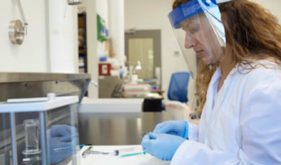
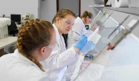
Having a well-trained team of histotechnicians is important in preparing the highest quality slides. StageBio employs technical trainers who are highly skilled histotechnologists and oversee the training of all laboratory personnel.
Our team routinely prepares upwards of 50,000 prepared slides each month - which requires a large dedicated and skilled team of histotechnicians. Each tissue leaving StageBio has been reviewed microscopically to ensure it is consistently placed, free of artifacts, and has crisp staining.
We offer:
Our histology services and facilities are GLP-compliant.
REQUEST A CONSULTATION
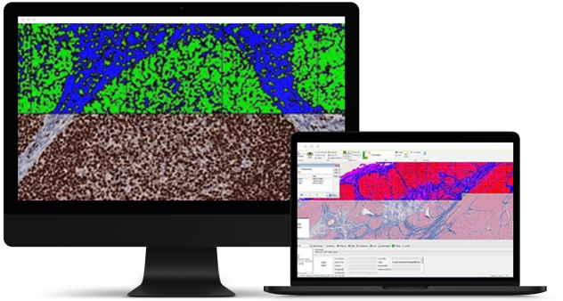
Image analysis of stained slides is a powerful tool that can add value to your drug development studies. Here at StageBio, we perform image analysis using high-quality scanned images.
We employ Visiopharm’s Biotopix™ solution and other image analysis solutions to extract quantifiable data from your stained slides. Image analysis can be used for the following common requests, as well as many more: linear and area measurements, counting positive cells and quantification of positive staining of proteins in cells and tissues. Here are just a few examples of the kinds of image analysis that StageBio can provide for you:
Data can be provided in Microsoft Excel format or in graph form. We can also provide statistical analyses and histopathological interpretation of the data.
To request a consult, click on the link below to start the process. We look forward to hearing from you!
REQUEST A CONSULTATION
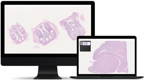
Converting glass microscope slides to high resolution digital images allows clients to view the slides we prepared from any computer with ease. Digital slides can be viewed online via StageBio’s image management software, or we can provide the images to you electronically.
Our high-capacity whole slide scanners can scan slides at 20X and 40X magnifications. Brightfield and multiplex fluorescence scanning capabilities (up to 6 channels) are available and we can scan both single (1x3) and double-width (2x3) slides. These images can be accessed at your convenience with our image-sharing service, which is very convenient for pathology discussions and internal review, without the need to ship the slides.
To request a consult, click on the link below to start the process. We look forward to hearing from you!
REQUEST A CONSULTATIONYes - StageBio has a staff courier and can arrange to pick up your study materials no matter the size or location – including internationally.
Place your tissue(s) in 10% neutral buffered formalin for between 24 and 48 hours, then place in 70% ethyl alcohol.
Access and complete the Contact Us form within this website to set up a facility inspection.


Director of Pathology
Contract Research Organization
Assistant Director of Medical Devices
Medical Device CRO