StageBio provides tissue cross-reactivity (TRC) studies for monoclonal antibodies and related antibody-based therapeutics, helping drug developers characterize intended and unexpected binding across human tissues.
From early feasibility and IHC method establishment through validation, GLP tissue screening, pathology evaluation and final reporting, our scientists and pathologists develop a robust study strategy, protect timelines and produce clear, defensible results.
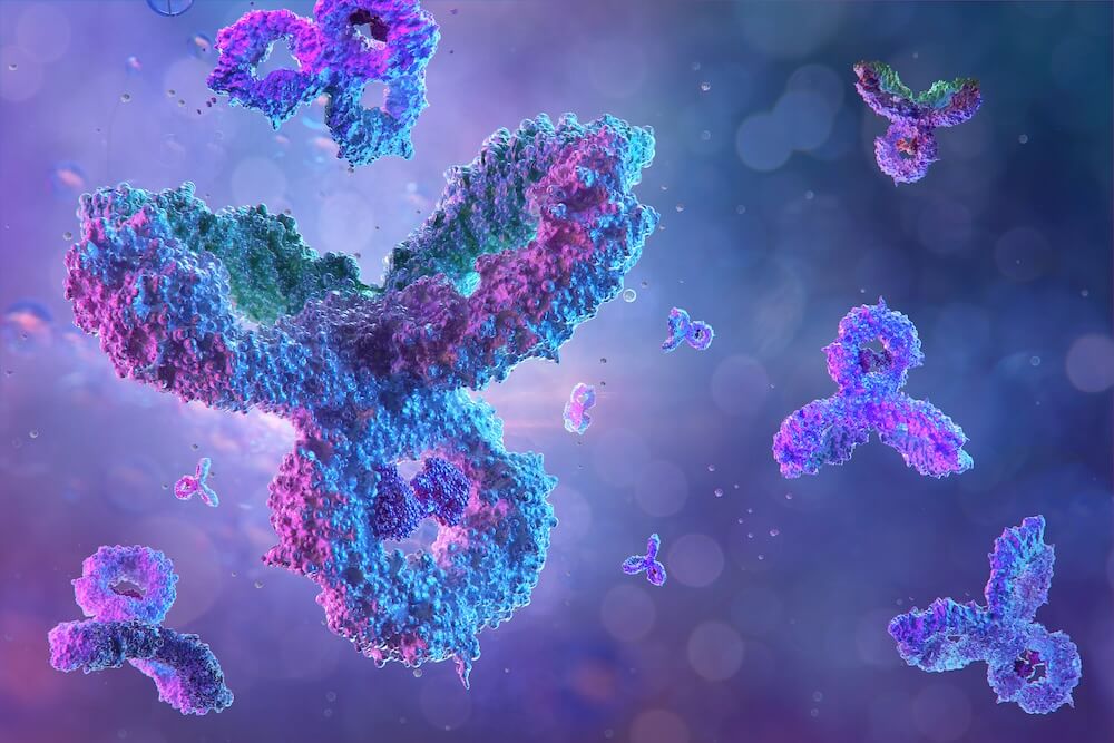
TCR findings feed directly into your nonclinical safety package and your IND, CTA or first-in-human timeline. ICH S6(R1) identifies human-tissue TCR as a recommended component for many monoclonal antibodies and related biologics. Planning early gives you time to interpret and, if needed, address unexpected tissue binding before it becomes a late-stage holdup.
A well-designed TCR study can identify:
Expected binding associated with the intended target
Previously unrecognized sites of on-target binding
Unexpected or potentially off-target tissue binding
The specific cells, tissue structures, and subcellular locations showing staining
TCR provides information about the distribution and character of tissue binding. It does not, by itself, demonstrate that biological activity or toxicity will occur in vivo. Findings should be interpreted alongside:
Target biology and normal tissue expression
Binding affinity
Tissue accessibility
Pharmacological activity
Relevant-species data
General toxicology findings
Clinical dose and route of administration
This is why experienced pathology interpretation is essential. As part of every TCR study, StageBio’s board-certified pathologists evaluate the location, distribution, and intensity of staining, identify the cell types and anatomical structures involved, and help interpret the potential significance of expected and unexpected binding. Your team isn't left interpreting ambiguous findings alone.
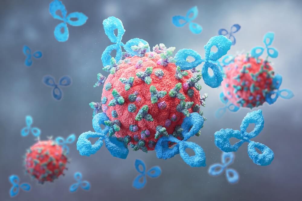
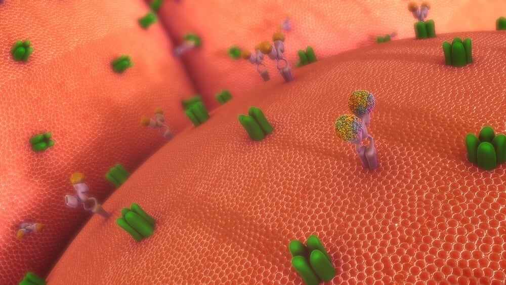
A validated method is the foundation for reliable, defensible results. Before the formal study begins, StageBio develops an automated IHC method tailored to the Test Item and matching Control Item.
Using positive and negative control tissues, we optimize concentration, detection chemistry, and staining conditions. This achieves clear, specific staining with minimal background. The method is then validated across three replicates in three independent runs to confirm repeatability and reproducibility. Removing antigen retrieval as these are run on fresh frozen tissues and this is not used.
StageBio can also transfer and validate an existing client method on our automated staining platforms.
Deliverables: Validated IHC protocol, selected study concentrations, defined control strategy, and method-establishment report.
Once the method is validated, it moves into the formal GLP study. The validated method is applied across three independent full human tissue panels representing the major organ systems.
Each tissue is evaluated using the Test Item at the selected high and optimized concentrations. The matching Control Item and appropriate positive and negative controls are run alongside it.
A board-certified pathologist assesses the location, distribution, intensity, and character of staining, including any unexpected or potentially off-target binding. Findings are provided in a comprehensive GLP report with representative images and tissue-by-tissue summary tables.
Selected animal tissues or panels may also be included where scientifically justified.
Deliverables: GLP tissue screening, pathology interpretation, representative images, findings tables, and regulatory-ready reporting.
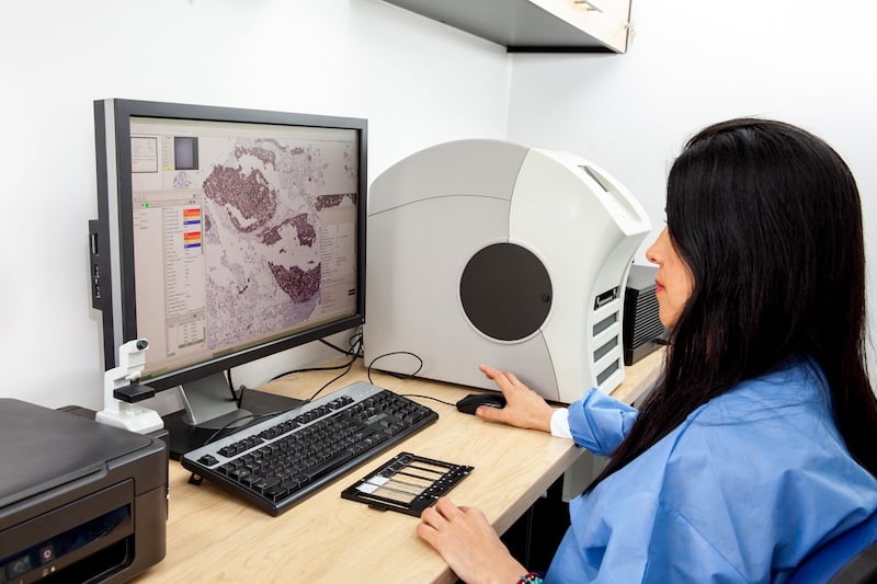
Unresolved tissue-binding questions can cost months if they surface close to an IND or CTA filing. Our TCR specialists and board-certified pathologists work alongside your team from early feasibility through final reporting, so findings are identified and interpreted well before they threaten your timeline.
7 Common Mistakes in Tissue Cross-Reactivity Study Planning (& How to Avoid Them)
What Is a Tissue Cross-Reactivity Study?
Use Cases and Benefits of Immunohistochemistry, Immunofluorescence, and In Situ Hybridization
A TCR study helps identify where an antibody-based therapeutic binds across human tissues. It can confirm expected binding, reveal previously unknown on-target distribution, and identify unexpected tissue binding that may require further interpretation.
No. The appropriate non-clinical strategy depends on the product, indication, development pathway, and available safety information.
ICH S6(R1) identifies human-tissue TCR as a recommended component supporting initial clinical dosing for monoclonal antibodies and related products. However, ICH S9 notes that TCR studies generally have limited utility for some advanced-cancer programs unless there is a specific concern or no pharmacologically relevant species is available. The requirement should therefore be assessed on a program-specific basis.
StageBio can work with your toxicology and regulatory teams to develop an appropriate study scope.
Planning should begin as early as possible once the clinical candidate and intended development timeline are understood.
Based on StageBio's current indicative timelines, sponsors should allow approximately 20 weeks. This covers method establishment and validation, the main study, and pathology reporting. Additional time may be needed for conjugation, material shipment, control sourcing, contracting, or method troubleshooting.
Not necessarily. Ex-vivo tissue binding does not automatically demonstrate biological activity or toxicity in vivo.
Several factors should be considered before determining the potential safety significance. These include the tissue structure involved, subcellular location, accessibility of the binding site, staining intensity, target biology, dose, pharmacology, and relevant species data.
A matching control helps distinguish test-item-specific staining from background, detection-system effects, and non-specific binding.
The most appropriate control should reflect the relevant structure or backbone of the clinical candidate as closely as possible.
Some clinical candidates do not perform effectively as IHC reagents. This may be due to insufficient tissue binding under assay conditions, detection limitations, high background, or technical incompatibility.
Early feasibility and method-establishment work help determine whether an interpretable method can be developed. ICH S6(R1) recognizes that a TCR study may not always be technically feasible when the clinical candidate is not a suitable IHC reagent.
StageBio has access to human and species-relevant tissue resources. Human tissue panels form the primary focus of the standard TCR approach.
Selected animal tissues can be considered where there is a specific scientific rationale, such as investigating unexpected human-tissue binding or supporting a wider species-relevance assessment.
Yes. StageBio can review an existing method and determine whether it can be transferred or adapted to our staining platforms.
Transfer requirements will depend on the method, detection system, controls, tissue preparation, and existing validation data.
Conjugation requirements should be discussed during the initial technical review. StageBio can advise on suitable test and control-item formats and coordinate with external partners where appropriate.
A post-conjugation binding assessment may be recommended to confirm that conjugation has not adversely affected target recognition.
The precise panel is defined in the study protocol and aligned with the therapeutic product, applicable guidance, and regulatory strategy.
StageBio maintains access to snap-frozen human tissue panels and selected animal tissues. Tissue suitability and antigen preservation are considered during study planning.
A pathologist records the structures and cell populations stained, along with the location, distribution, intensity, and character of the staining.
Findings are compared with the matching control and interpreted in relation to known target distribution and the wider development program.
Yes. StageBio can provide whole-slide scanning and secure digital access to support sponsor review, pathology collaboration, and project discussions.
Brightfield slides can be scanned at appropriate magnifications according to the study requirement.
TCR is not generally recommended as a comparability assay because it may not detect subtle changes in critical quality attributes following manufacturing-process changes. Other analytical and functional methods are usually more appropriate.


 513.204.4400


