Successful preclinical research programs rely on board-certified veterinary pathologists to provide critical and accurate analyses pertaining to safety and clinical relevance of the experimental product. To obtain the most comprehensive and high-quality final reports, it is vital to ensure that the study pathologist has access to the most effective support team and most pertinent tools available.
Below is a list of items that will provide crucial support to the study pathologist and will ensure the highest quality pathology report:
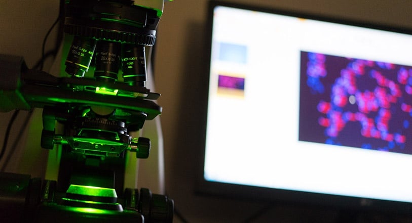
1. Microscope and Camera
At the heart of a pathology evaluation is the microscopy equipment and slide quality on which the assessment is made. As an example, a hematoxylin and eosin (H&E) stain analyzed by light microscopy is the gold standard for most histopathology work. It gives an overview of the tissue, the results are reliable, and pathologic abnormalities are easily identifiable by an experienced pathologist. Although techniques like these are routine in the histology lab, the slide quality and microscope capability need to meet high standards to ensure accurate analysis.
While years of training and experience are invaluable, ultimately the study pathologist will be making decisions about pathologic changes in the tissue based on light or fluorescence microscopy. The quality of the tools used to capture images has bearing on the ability of the pathologist to capture changes within a tissue, which is of the utmost importance. It is therefore critical that the slides prepared and methods used are reproducible between samples and experiments, and that the technology being employed is maintained annually (or semi-annually) and in good working order. Only these rigorous measures allow for accurate analysis of histopathological data.
Quantitative analysis of high-resolution images is also a powerful tool, allowing the user to set thresholds and generate measurements from a tissue section. Guided by the expertise of a veterinary pathologist or experienced morphometrist, the computer program can acquire, import, process and analyze an image from a slide placed on the microscope stage. For example, in one section it is possible to record and quantitate:
- Number of cells stained with a marker
- Length/area of a lesion or other structure within a given tissue
- Pattern or intensity of a particular immunohistochemical stain
- Ratio of one cell type versus another
Considering the laboratory and animal costs associated with conducting preclinical studies, it is vital that as much data is collected from each slide as possible. The most advanced microscopy equipment ensures that images can be captured at high resolution, providing a detailed and instrumental window into the histopathology underlying the study.
2. Research Associate
Working with a research associate (RA) can help contract laboratories increase productivity while maintaining high-volume capacity. RAs provide technical and administrative support to veterinary pathologists who can consequently focus their expertise on making detailed histopathological assessments and producing high quality pathology reports. A capable RA will help QC pathology data and maintain timelines by coordinating study activities with other company personnel. Employing an RA as a support person will help ensure that all aspects of the study, including data collection, analysis and report writing are done according to protocol.
3. Quality Assurance Unit
A dedicated team responsible for auditing all study data and results, and for overseeing quality and compliance are critical when working with a GLP study. The quality assurance unit (QAU) will identify problems and correct them at all stages in the study including: study set up, slide preparation, pathologic evaluation and long term archiving. This level of auditing provides confidence to the laboratory staff and clients that the highest quality data collection and sample analysis is being conducted.
4. High Quality Slides
The quality of the necropsy as well as the prepared microscope slides have a major impact on all other aspects of the final report. High-resolution images of low-quality slides, for example, will not be conducive for providing usable data to the sponsor. Reliable methods and experienced laboratory staff are essential to ensure slides are prepared properly and consistently. Procedures must be verified and followed carefully. From tissue collection to sectioning and staining, all solutions and equipment must be of the highest quality and all technical staff must be well trained. An efficient and experienced laboratory staff will support the veterinary pathologist allowing him or her to prepare a solid pathology report.
In the era of digital pathology, another important consideration for slide preparation is the ability of the lab to convert glass sides into whole slide images for storage, sharing and further analysis. Digital pathology allows for more advanced image analysis and quantitation as whole slides are scanned using a whole slide scanner then analyzed on a high-resolution screen. It also facilitates more efficient data management and collaboration.
5. Data Capture System
Effective data capture and management is essential when collecting, processing and storing large amounts of critical preclinical data. A successful system should be fully integrated and capable of providing automated, reliable support at each step. From breeding and animal tracking, to image collection and analysis, to final study submission; the properly implemented data capture system should improve workflow, ensure compliance, support data quality, and be validated!
Remember: an experienced, well-equipped and supported veterinary pathologist will provide the highest quality reports for your preclinical research program.