

Histomorphometry is a valuable tool when used in conjunction with histopathological evaluation to assess the healing of bioresorbable or biostable orthopedic implants.
The orthopedic medical device industry has been expanding rapidly in the last few years. New advancements in device types, more advanced materials and biomaterials are changing the way these devices are used. Orthopedic implant studies are complex and nuanced, leading to our discussion here of histomorphometry.
Here, we'll review a portion of this 2019 article: Histopathological Evaluation of Orthopedic Medical Devices: The State-of-the-art in Animal Models, Imaging, and Histomorphometry Techniques by Nicolette Jackson, Michel Assad, Derick Vollmer, James Stanley and Madeleine Chagnon.
What Is Histomorphometry?
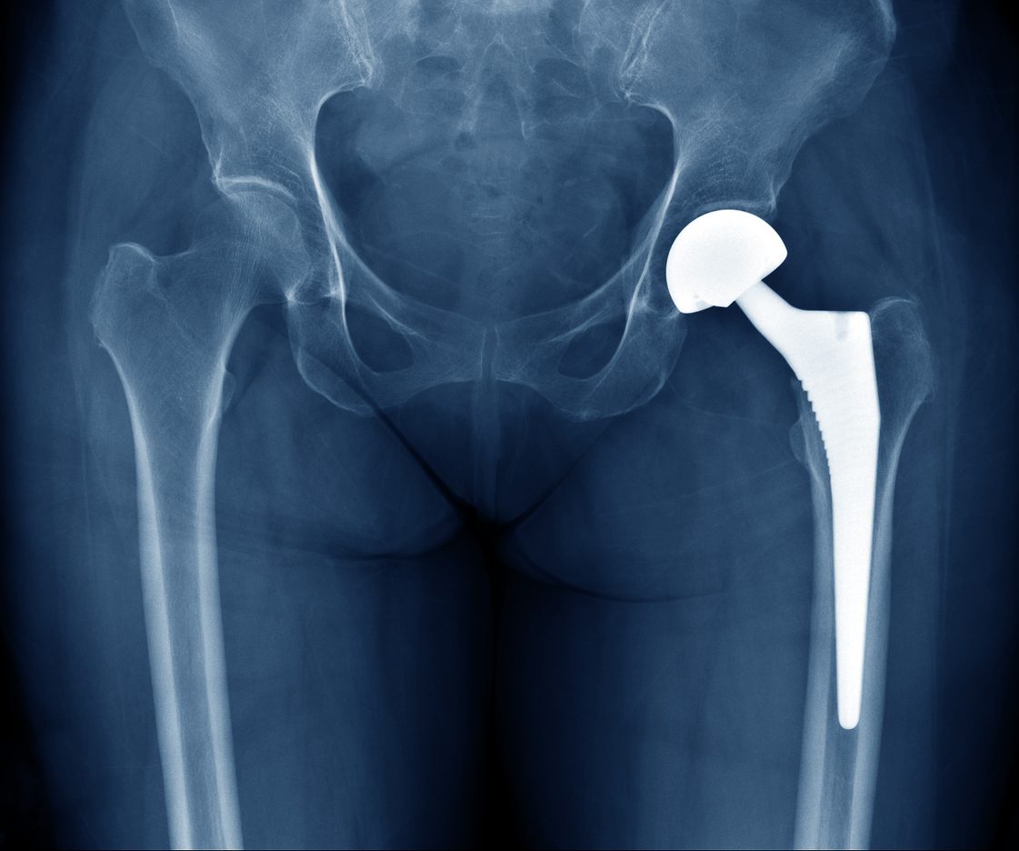
Histomorphometry is described as the measurement of various lengths, areas, shapes, or regions of interest in a tissue. In an orthopedic context, histomorphometry provides valuable information about the amount of bone-implant contact (BIC, osseointegration) with a biostable device and can demonstrate the amount of new bone growth into a region of interest (ROI), as well as residual implant material and fibrous connective tissue
Per Jackson et al, histomorphometry can be divided into static or dynamic morphometry techniques.
Both techniques require the use of an appropriate slide scanning system to image the entire histological ROI. The captured image is then utilized for morphometry analysis techniques with an image analysis program.
Notes on Static Morphometry
Possibly the most important parameter to analyze for biostable implants is bone-implant contact (BIC). It is measured by tracing the edge of a solid implant to obtain the entire perimeter, and additionally tracing areas that demonstrate osseointegration (BIC) with the implant surface. The result is presented as a percentage.
Certain parameters within the ROI can usually be color segmented. For instance, new bone formation and fibrous connective tissues can often be segmented, though this, depends on the histological appearance and composition of the implant material.
Note that in some situations, implanted biomaterials will stain very similarly to bone, so color segmentation may not differentiate the bone growth from implant material. In these situations, the implant material may need to be hand-traced (a time-consuming task). Trying different bone stains may solve the issue, but usually the implant will stain similarly to bone regardless of the histological stain that is used. In a nutshell, the implant will be visible to humans but not to a majority of computer programs, though advanced artificial intelligence (AI) techniques may prove useful for conducting histomorphometry on these types of materials in the future.
Notes on Dynamic Morphometry
Dynamic morphometry is used to demonstrate the rate of new bone growth and mineralization. Throughout the in-life phase of the study, the animal is treated with various fluorochrome labels (such as xylenol orange or calcein blue) at predetermined intervals (like every two weeks, for instance). Immediately upon euthanasia, tissues are harvested, fixed, and typically ground MMA sections are made.
Unstained slides are scanned with an immunofluorescence scanner and those images are used for dynamic morphometry measurements. Once scans and analyses are confirmed to be acceptable, the original slides can then be stained and utilized for the histopathology and static histomorphometry analyses.
Goals
A typical goal for dynamic morphometry is to measure the rate of bone mineralization by measuring the distance between various colors of fluorochrome labels.
Note that the route of administration of the fluorochrome will affect the results, so proper planning of dosing concentrations, dose timing, and histological section thickness is required to optimize the visualization of fluorochrome labelling in the images.
Conclusion
To summarize, static histomorphometry allows us to study the effects of orthopedic implants on bone and surrounding tissue at the time of necropsy; while dynamic morphometry allows us to visualize the bone growth process, over a longer period of time. If you'd like to learn more about histomorphometry services visit our website or contact us!
