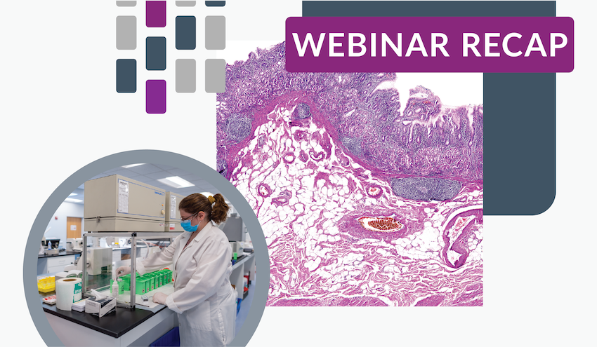


We recently hit the webinar stage to give an in-depth look at what it takes to maximize the value of your high-throughput histopathology study. Our goal was to not only outline the basic techniques and philosophy behind histopathology, but to also demystify the entire process.
In our humble opinion, the webinar was a success. (And that’s in no small part thanks to everyone who attended!) But if you wanted to join us but couldn’t make it, don’t worry; we’ve written this blog to highlight a few of our webinar’s key takeaways. Keep reading for insight into what a high-throughput histopathology lab does—and how you can make your next histopathology study a success.
What is the purpose of histopathology in preclinical research?
First, let’s break down the goal of the histopathology study phase into three parts:
- Identify and characterize test article-related changes in tissues and organs
- Decide their pathologic importance to the overall health of the animal
- Predict their relevance to humans
Looking at the overarching goal, it’s easy to see that histopathology is an important component of toxicity testing and efficacy studies. In other settings, histopathology may be used to characterize the phenotype of experimental or genetically engineered models or to determine the cause of illness or death in a diagnostic pathology case.
How does the histopathology phase begin?
The histopathology phase of a research study really starts during study design and protocol development. During this time, clients (investigators) provide the draft study protocol for technical review by the Laboratory Director and Pathology Principal Investigator to ensure the histopathology section is well defined and has a clear directive.
Once a study protocol has been finalized, it’s important to schedule slide preparation (histology) and tissue evaluation (histopathology) quickly with a pathology service provider like StageBio to keep the study on its intended timeline. There is a finite amount of bandwidth within a histology laboratory, and the schedules of pathologists can fill up quickly in today’s research climate. Don’t get caught in a backlog!
To maintain the integrity of the final pathology report, it is critical to maintain excellence throughout the entire process, beginning with tissue collection. In other words, quality starts at necropsy with trained, skilled prosectors. These prosectors must harvest tissues consistently to allow for thorough fixation while not damaging them during handling and weighing. Prosectors must also properly document and collect gross lesions noted at the time of sacrifice. These necropsy findings should be communicated to the histology laboratory prior to the start of tissue trimming so they can be recorded on histology paperwork, captured on slides, and ultimately correlated with microscopic findings by the Study Pathologist and included in the final report.
At StageBio, we receive study materials in one of two ways:
- Our clients ship us fixed tissues in bags or jars of formalin (or their fixative of choice).
- Our courier comes to a client’s facility and picks up the tissues and other study materials.
Sending a courier is generally the safest and most efficient way to ensure your study materials arrive on time to the histology laboratory.
Once preserved tissues are received, specimens are carefully inventoried before commencing any laboratory procedures. All technicians who will be involved in the histology phase review the study protocol so they are prepared to handle and process each protocol-required tissue.
The histology laboratory must trim, embed, section, and stain the tissues from each animal as consistently as possible. Tissues are trimmed in an anatomically standard orientation and placed in cassettes with indelible study and animal identification for easy tracking. The cassettes are then loaded into automatic processors, infiltrated with paraffin wax, and embedded into paraffin blocks.
Wax blocks containing protocol-required tissues are sectioned at 4 to 5-micron thickness using a microtome and affixed to glass microscope slides. Once the tissues have adhered to the slides, usually an overnight process, the slides are stained with routine hematoxylin and eosin (H&E) stain. Other special stains may be used depending on study requirements at the request of the Study Sponsor, Study Director, or Study Pathologist.
Once the slides for each animal have been stained and cover slipped, each slide is matched to the block to ensure proper identification. Lastly, quality control technicians review every single section on every single slide to check that each tissue is present, and that the staining is crisp and free of any artifacts.
Now, slides are ready for microscopic evaluation.
How is histopathology data captured?
It’s easy for outsiders to think that a “black box” exists between the study tissue specimens affixed to glass slides and the histopathology data recorded into an electronic data capture system or spreadsheet. Slides arrive on a pathologist’s desk and, as if by magic, data appears on the back end. But what’s happening when the slide is under the microscope, being analyzed by the pathologist?
We’ll need to answer a few questions to help provide context:
What does an anatomic pathologist do?
An anatomic pathologist performs careful pattern recognition, scanning tissues at the microscopic level for changes in tissue architecture, infiltrating cell types, and altered appearance of cells that compose the tissues. These patterns could indicate changes in cell function or viability. Even control animal tissues have a range of subtle morphologic differences considered normal for the strain, species, and age, so a lesion is considered a change that is outside of that range.
Experienced pathologists have a skill set that can be tailored by research setting, recording and interpreting study data to best answer the question being asked:
- In diagnostic cases, splitting out fine details of microscopic changes and attributing significance to each one
- In toxicology studies, remaining consistent in lesion terminology and setting appropriate thresholds
- In novel experimental models, recording even subtle changes in anatomy and recognizing the normal background appearance of tissues in a particular animal strain
If you are an investigator, remember that the pathologist is your partner! Helpful ancillary information to provide before slide evaluation includes study design, test article mechanisms, findings in previous studies using the same test article or animal model, in-life findings, clinical pathology data (including hematology, clinical chemistry, urinalysis, coagulation data, hormone analysis), organ weights, and necropsy observations. This information helps to provide the most meaningful interpretation of microscopic findings.
When a pathologist scans slides for lesions, what is an appropriate threshold for normal vs. abnormal?
Even in tissues from control animals, it’s common to find pathologic processes at work. These are often subtle and functionally inconsequential tissue changes but occasionally meaningful to the life and well-being of the animal.
Many non-neoplastic microscopic findings exist on a spectrum with no firm line between affected and unaffected animals. With increasing experience, every pathologist refines their mental image of the typical “normal” appearance of a tissue. This mental image, backed by experience, informs a pathologist’s minimum threshold for recording a pathologic change. For example, a cluster of lymphocytes in the lacrimal gland of a rat or a single degenerative tubule in the kidney of a mouse might be considered “below threshold” by some pathologists, which means it’s within the range of normal for the age and species and not worth recording in the pathology data.
In a semi-quantitative histopathology assessment, which is used when broadly surveying tissues for changes and grading them on a 0-4 or 0-5 severity scale, a minimum threshold for recording a tissue abnormality is used. Too low of a threshold can result in overly complex data and spurious—or chance—differences in the incidence of trivial background findings across treatment groups. Conversely, too high of a threshold may result in missed treatment-related differences in incidence or severity of lesions, especially those that are of similar character to background changes. An appropriate threshold finds a balance.
There is some wiggle room, however, when setting appropriate thresholds. One pathologist might use a slightly lower threshold for recording a microscopic lesion than another, yet both pathologists detect a test article-related change in the incidence and severity of that lesion across study groups despite the small difference in threshold that shifts the overall severity downward or upward.
How important is lesion terminology?
Terminology is key to effectively communicating study findings. Often, multiple terms can be used to accurately describe the same gross or microscopic lesion. We recommend that each testing facility develop a list of accepted descriptive terms for necropsy technicians to utilize to maintain consistent gross lesion terminology within and between studies.
At microscopic evaluation, there are virtually unlimited options to describe tissue changes. Terminology selection can impact comparison between studies of similar test articles or animal models, as reader confusion may result if different terms have been used to describe the same lesion. What might be “mixed cell infiltrate” to one pathologist could be “chronic active inflammation” to another pathologist.
The ultimate decision of which term to be used for a microscopic lesion lies at the intersection of four factors:
- the pathologist’s professional assessment of the term that best fits
- study-specific terminology as requested by the investigator and/or based on previous studies or publications
- consideration of globally accepted terms in harmonization documents
- regulatory agency requirements
Are there any standardization efforts in lesion terminology?
A movement over the past two decades has made progress in harmonizing the terminology used by toxicologic pathologists. A global project called The International Harmonization of Toxicologic Pathology Nomenclature (INHAND) has produced 19 guides to date through the collaboration of professional organizations, including Society of Toxicologic Pathology (STP) in the United States. These guides are organized by organ system and species, and they define common lesions with example photomicrographs.
Other published resources exist that intersect meaningfully with INHAND guides include:
- The National Toxicology Program Non-Neoplastic Lesion Atlas
- Standardized System of Nomenclature and Diagnostic Criteria (SSNDC)
- GoRENI/Registry of Industrial Toxicology Animal-data (RITA)
- Standard for Exchange of Non-Clinical Data (SEND)
Standard for Exchange of Non-clinical Data (known as SEND) is a way of presenting data in a consistent format for Investigational New Drug (IND) and Biologic License Application (BLA) submissions to the US Food and Drug Administration (FDA). To make histopathology data fit into a SEND format, toxicologic pathologists try to use approved terminology (for morphologies, modifiers, and tissue names) that is available as a large but sortable document and regularly updated on the CDISC website. There is intentionally a high level of overlap between SEND approved terminology and INHAND guides. If a non-SEND approved term is used, and this term hasn’t been “mapped” to a SEND-approved term on the back end, that term will be flagged when the data is prepared for submission. Keep in mind that SEND mapping does not alter the original data as captured during the histopathology evaluation, and this original data also remains available to regulators.
*******
So, to collect accurate and reliable histopathology data, there’s a lot going on behind the scenes—from the histology process to the pathologist’s microscopic evaluation to the clear presentation of results in a pathology report. And with so many variables and moving components, that makes your choice of which histopathology team to partner with incredibly vital.
How StageBio can help investigators with high-quality, insightful histopathology
At StageBio, we utilize our expert team and infrastructure to provide quality results with a reasonable turnaround time. In the project planning phase, we work efficiently with investigators to identify histopathology needs. We then bring in experts across our client services, histology, and pathology teams to ensure that the end result will help to answer your scientific questions at hand.
We thoughtfully plan out the histology process so your pathology evaluation is set up for success from the start. We have two high-throughput laboratories staffed with skilled technicians who routinely create approximately 1,000 slides per day. These technicians are experienced in routine and custom trimming, embedding, and sectioning with automated staining. And while they prepare a massive number of slides, they do so while maintaining the highest quality and consistency.
Our pathology team consists of approximately 30 board-certified veterinary pathologists, with the general toxicologic pathology division making up half of the group. The experience level of this team is notably high, with many of our pathologists having 10 to 30 years in the field of toxicologic pathology, including positions of leadership. Our pathologists can advise on all parts of the study process and interpret pathology findings in context with in-life and clinical pathology data, providing an integrated interpretation of macroscopic and microscopic findings.
Conclusion
To learn more about how to set up your high-throughput histopathology study for success, contact our team here.
