

IHC is a valuable tool that can be used during most phases of drug development. It assists with target validation during early discovery projects and can help determine efficacy and safety during later phases of research and development.
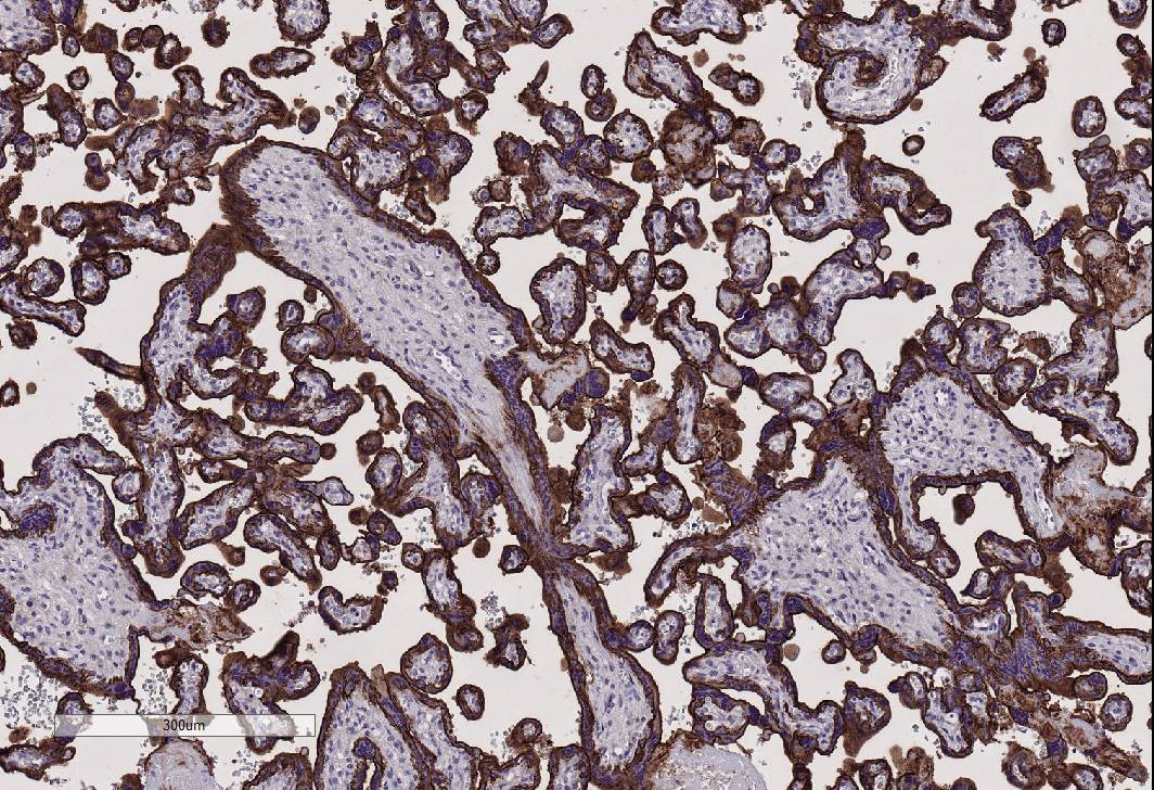
Unlike other antibody assays, IHC allows researchers to visualize exactly where proteins are localized within tissues (specific cell types and subcellular localization; cell membrane, cytoplasm or nuclear).
Examples of how IHC can be used in both GLP and non-GLP preclinical studies1:
- Analysis of Rodent Carcinogenicity studies1:
- Use of IHC to determine lineage of neoplasms that are identified in routine H&E sections.
- Cytokeratin-positive tumors are of epithelial lineage, Vimentin-positive tumors are of mesenchymal lineage and CD45-positive tumors are of lymphoid lineage.
- Additional markers may be used to further identify tumor origin.
- Markers, such as Ki67 or PCNA can be used to determine how rapidly neoplastic cells are proliferating.
- IHC can also be used to distinguish apoptosis from necrosis.
- Endothelial cell markers, such as CD31, can be used to evaluate tumor angiogenesis.
- Use of IHC to determine lineage of neoplasms that are identified in routine H&E sections.
- Tissue Cross-Reactivity Studies1:
- IHC provides the ability to map the presence of targets across many tissues with the use of Tissue Micro Arrays (TMAs).
- Potential off-target toxicity can be identified by staining TMAs with candidate antibodies.
- IHC using potential therapeutic monoclonal antibodies can be used to identify target and off-target binding of antibody in human tissues.
- Biodistribution Studies1:
- IHC using client-provided therapeutic monoclonal antibodies can help identify differences between animal models and humans in terms of localization and binding of specific targeted epitopes.
- Use of IHC to Identify and Characterize Biomarkers1:
- IHC can be used to identify biomarkers of cell death (e.g., caspases), toxic injury (e.g., peroxisome markers) and markers of disease (e.g., beta amyloid in Alzheimer’s, insulin in diabetes).
- Tumor Microenvironment of Xenografts:
- IHC can be used to identify and classify inflammatory markers such as CD3 and other T-cell subsets as well as macrophages and neutrophils, among others.
- IHC can also be used to identify tumor markers such as PD1, PDL1 and EGFR.
- Identification of human stem cells in animal tissue:
- IHC can be used to distinguish human from non-human cells in animal tissue.
- Some specific IHC markers which can be used include; human mitochondria and human nuclear antigen.
Ideally, it is best to identify IHC markers of interest in the planning stage of a study. Once IHC markers have been identified, the appropriate methods for tissue collection and fixation can be determined. Most antibodies will work on formalin-fixed, paraffin-embedded tissues, but some will only work on frozen tissues.
While many commercially available antibodies have been validated for both mouse and human tissues, the majority of IHC antibodies on the market are made specifically against human proteins. When selecting an antibody for IHC, it is critical to do a thorough literature review of the target and available antibodies for IHC in order to determine species cross-reactivity. In some cases, available antibodies will not have been validated in the species of interest or in the tissues being evaluated requiring validation of the antibody in this species for a particular tissue.
HSRL’s highly experienced team is proud to offer a broad range of routine and advanced IHC and IF staining techniques for both frozen and FFPE tissues. Numerous antibodies across multiple species have been validated in-house, including mouse, rat, dog, pig, non-human primate and human. We offer single and multiplex chromogenic and immunofluorescent IHC staining for all of your protein localization and co-localization studies.
HSRL has the ability to perform traditional manual staining as well as automated IHC using the Biocare Intellipath FLX IHC Stainer. The Biocare stainer is an excellent system for research applications and efficiently facilitates large batch slide runs. Automation allows for increased throughput and consistent results for larger studies.
Our highly trained team of scientists can assist with your research goals by providing guidance regarding which IHC markers and antibodies will be most effective for your studies. We are a fully GLP-compliant laboratory with extensive experience with both GLP and non-GLP IHC method development and will optimize the antibodies that fit your research needs. We are happy to consult with you regarding recommendations for tissue collection and fixation procedures that will lead to the best possible staining results.
References:
- Oyejide L., Mendes O., and Mikaelian I. (2016).Molecular Pathology: Applications in Nonclinical Drug Development. In A. S. Gaqi (Ed), A Comprehensive Guide to Toxicology in Nonclinical Drug Development (Second Edition) (pp. 237-276). Washington, DC: Academic Press.