

The ability to convert glass microscope slides to high resolution, digital images and then perform morphometric image analysis is an important part of many preclinical research and development studies.
HSRL uses a Hamamatsu Nanozoomer S60 high-capacity whole slide scanner, which can scan slides at 20X and 40X magnifications. It has Brightfield and multiplex fluorescence scanning capabilities (up to 6 channels) and can scan single-width (1x3) and double-width (2x3) slides.
Whole slide scanning has several advantages:
- Allows multiple people to view slides at once during conferences and presentations
- Provides a permanent record of IF-stained slides, which fade rapidly losing critical data
- Offers the ability to view all channels of multiplexed IF-stained slides at once
- Images can be uploaded onto a server, such as ShareFile, which allows clients to download and view them for examination, publication and analysis
- Whole slide scans provide the best platform for morphometric analysis since all sections on a microscope slide can be analyzed at once
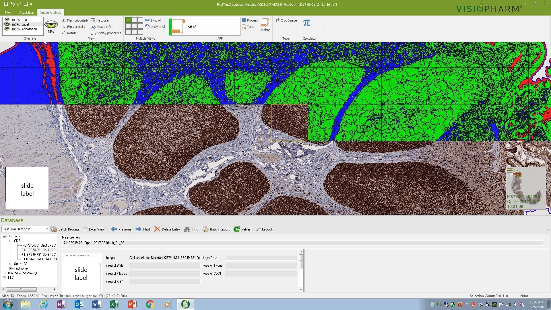
Morphometric image analysis of whole slide scans is a powerful tool that adds meaningful data to drug development studies. Morphometry can be used to perform linear as well as area measurements, count positive cells and provide quantifications of positive staining of proteins in cells and tissues, among other things. Here at HSRL, we use both the Visiopharm® Biotopix™ and Image-Pro Premier as our image analysis software.
Below are some examples of morphometric analysis that can be performed on scanned slides:
- Quantification of cell types and cancer biomarkers in tumor microenvironment studies (xenografts)
- Quantification and distribution of human stem cells in animal tissue
- Quantification of therapeutic monoclonal antibodies in biodistribution studies
- CD31 quantification for angiogenesis studies
- Ki67 or BRDU cell counts for proliferation studies
- TUNEL or Caspase-3-positive cell counts for apoptosis/programmed cell death
- Immunophenotyping; single or multiple immune cell markers
- GFAP and Beta-amyloid quantification in Alzheimer’s studies
- PAS-positive goblet cell counts and area in eyes
- Insulin and glucagon quantification in diabetes studies
- Measurement of wound healing/re-epithelization on skin
- Fibrosis quantification in muscle and liver
- Prussian Blue stain for iron quantification in liver
Morphometric analysis data can be provided as an excel table or a graph. We can also provide statistical analysis and histopathological interpretation of morphometry data.
To request a consult, contact us to start the process. We look forward to hearing from you!