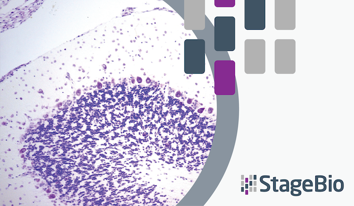
Learn what triggers minimal Apoptosis/Single Cell Necrosis in nonhuman primate brain in this whitepaper coauthored by StageBio senior pathologist Dr. Joy Gary.
Evaluating biodistribution and toxicity of biological-based test articles (such as AAVs and oligonucleotides)in nonhuman primates often requires extensive sampling for both molecular and histopathologic assays. To preserve the integrity of the nucleic acids of these test articles, researchers commonly use intravenous perfusion of ice-cold (2°C-8°C) saline at necropsy before fixing tissue for microscopic evaluation.
In recent preclinical studies evaluating biological-based test articles, minimal apoptosis/single cell necrosis (A/SCN) within the brain was observed, including in random neural cells throughout the cerebral cortex and, less frequently, the hippocampus across all animal groups (including controls).
Senior Pathologist Joy Gary, DVM, PhD, DACVP and co-author Brad Bolon, DVM, MS, PhD published a 2024 paper that explores one hypothetical explanation for the observable A/SCN: Ice-cold perfusion, in conjunction with tissue acidity and hypoxia, triggers A/SCN in the stressed cells. Other possible incidental or procedural causes for this finding were discussed including background apoptosis and pre-necropsy sedation.
As detailed in the paper, the cells affected by A/SCN exhibit cytoplasmic hypereosinophilia and other end-stage features. However, glial reactions, leukocyte infiltration/inflammation, or other parenchymal change are not present. The severity of this A/SCN is minimal for controls, but it could reach mildly exacerbated levels in treated samples.
Read the 2024 paper to learn why the authors suggest that A/SCN observed in nonhuman primate brains processed by ice-cold saline profusion with delayed immersion fixation is not a lesion related to test articles, but rather a procedural artifact.
