

Historically, success in pinpointing the microenvironmental contributions to soft tissue sarcoma progression, especially during the onset of sarcoma, has proven elusive. While animal models are used to try and uncover these microenvironmental contributions, researchers still find difficulty when attempting to:
- Discriminate between microenvironmental, precancerous, and cancer cells
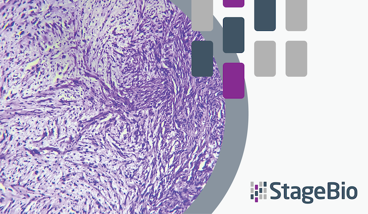
- Define a precancerous microenvironment during soft tissue sarcoma onset and progression, particularly malignant peripheral nerve sheath tumors (MPNST)
StageBio pathologist Olivia M. Patania, DVM, PhD, DACVP, participated in a study that sought to address this difficulty. To do so, the researchers developed a zebrafish model that allows for the segregation of these different cell populations using fluorescence-activated cell sorting, enabling detailed profiling and analysis of the microenvironmental contributions to sarcoma progression.
The researchers released their study’s findings in this paper titled, “Profiling the cancer-prone microenvironment in a zebrafish model for MPNST.” The study provides insights into how the microenvironment may regulate MPNST initiation and progression, highlighting potential therapeutic targets.
How the zebrafish model helps in studying microenvironment contributions to MPNST
The zebrafish model provided Olivia and her co-authors a system that allows for the segregation and analysis of microenvironmental, precancerous, and cancerous cell populations. Key features of the model the researchers developed include:
- Genetic Mutations: The zebrafish are genetically modified with mutations in the tumor suppressor genes TP53 and BRCA2, which predispose them to developing MPNST, mimicking the genetic conditions that can lead to this type of cancer in humans.
- Fluorescence-Activated Cell Sorting (FACS): The model uses a sox10:RFP reporter construct, which labels cancer and potential precancerous cells with red fluorescence. This allows researchers to use FACS to separate and analyze these different cell populations accurately.
- Predictable Tumor Development: MPNSTs in this model arise in a discrete anatomical location (the optic nerve pathway) within a predictable timeframe, facilitating the study of the transition from a precancerous to a cancerous state.
- Transcriptome Profiling: By using RNA sequencing (RNA-seq), the study profiles the gene expression of microenvironmental, precancerous, and cancer cells, providing insights into the molecular and genetic changes that occur during tumor initiation and progression.
- Comparative Genomics: The model allows for the comparison of gene expression changes between zebrafish and human MPNSTs, identifying conserved genetic contributors to cancer progression.
By introducing mutations in the TP53 and BRCA2 genes and using a sox10:RFP reporter construct, researchers were able to segregate and analyze microenvironmental, precancerous, and cancerous cell populations through FACS. The zebrafish model facilitates detailed transcriptome profiling via RNA sequencing, revealing significant activation of inflammation and immune-associated signaling networks. Comparative genomics between zebrafish and human MPNSTs identify conserved gene expression changes and potential therapeutic targets, such as the extracellular matrix protein periostin (POSTN).
Overall, the zebrafish model provides a powerful tool for understanding the microenvironmental factors that influence MPNST initiation and progression. This innovative approach provides critical insights into the role of the microenvironment in MPNST initiation and progression, offering new avenues for research and treatment strategies.
Access “Profiling the cancer-prone microenvironment in a zebrafish model for MPNST”
Read and download the full article from Oncogene here for more information on how the zebrafish model allows for a more comprehensive understanding of microenvironment contributions to MPNST initiation and progression
