

StageBio senior pathologist co-authors study on the use of 3D-printed bioactive scaffolds for restoring midfacial bone defects.
Complex critical-sized defects in midfacial bones
Critical-sized defects (CSDs) in midfacial bones are geometrically and mechanically complex, and they are often clinically challenging to treat. While autologous bone grafts are the gold standard, they are unable to fully replicate the complexity of the facial bone structure, resulting in a patient’s continued psychological distress and difficulties with vision, smell, speech, and mastication.
StageBio scientists, senior pathologist Dr. David Garlick and morphometrists Will Grundy and Derick Vollmer contributed to a paper highlighting the potential bone regenerative efficacy of using 3D-printed poly-and-caprolactone-decellularized bone (PCL-DCB) scaffolds augmented with stromal vascular fraction of cells from lipoaspirate tissue (SVF) to treat complex CSDs in the zygomatic bone.
Testing the clinical application of 3D-printed PCL-DCB scaffolds
To test the viability of 3D-printed PCL-DCB scaffolds augmented with SVF to treat CSDs in the zygomatic bone of humans, the study design relied on established findings that porcine facial bones mirror human facial bones in terms of density, cross-section, lamellar bone structure, and growth rates.
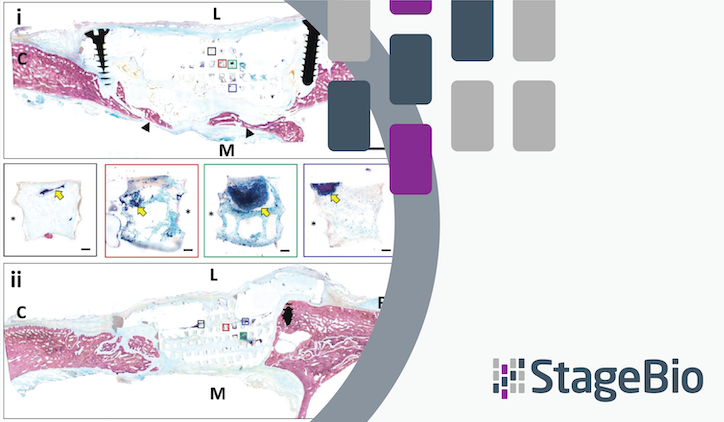
Scientists at Johns Hopkins University, including lead authors Drs. Srujan Singh and Warren Grayson, designed a point-of-care (POC) therapeutic strategy to treat defects that were 2 centimeters thick, segmental, and bilateral in the zygomatic arches of Yucatan minipigs with skeletally mature facial construction. These defects were created using animal-specific cutting guides according to pre-treatment CT scans. Afterward, the team treated the defects with 3D-printed PCL-DCB scaffolds augmented with autologous porcine SVF. The hypothesis was that this course of treatment would promote robust bone regeneration and facilitate an interoperative reconstruction strategy that is clinically translatable.
Creating the 3D-printed PCL-DCB scaffolds
A CT scan was conducted on each minipig to 3D print customized porous PCL-DCB scaffolds with solid fixation tabs. Subcutaneous adipose tissue was harvested from each minipig’s dorsal lumbar region, with the SVF being isolated and resuspended in fibrinogen. These cells were then injected into the pores of the 3D-printed scaffolds after they were implanted to treat the surgically created defects in the zygomatic arches.
An overview of the study results
The study’s findings are presented in the peer-reviewed article, “Point-of-care treatment of geometrically complex midfacial critical-sized bone defects with 3D-Printed scaffolds and autologous stromal vascular fraction,” which you can access here.
By reading the article, you can learn the details behind its findings, namely that 3D-printed PCL-DCB scaffolds:
- Restored zygoma anatomy
- Promoted bone regeneration and osseointegration
- Provided a template for deposition of mineralized bone nodules
Overall, the study concluded that 3D-printed PCL-DCB scaffolds augmented with SVF illustrate the viability of biomaterial implants with the correct biochemical and biophysical properties as a clinical solution that promotes complete healing of complex CSDs in midfacial bones.
Access the article, “Point-of-care treatment of geometrically complex midfacial critical-sized bone defects with 3D-Printed scaffolds and autologous stromal vascular fraction,” here.
.
